 Appointment Guide
 Poll
Poll

Read on to learn more about wet macular degeneration.
What are the causes and risk factors for wet macular degeneration?

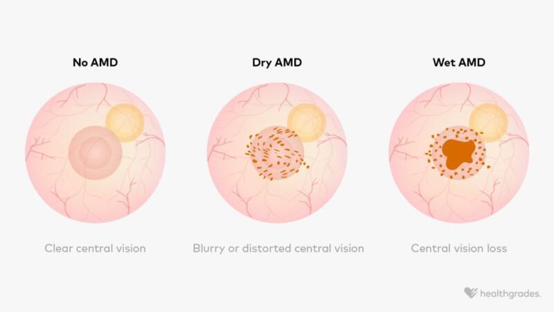
Wet AMD occurs when new blood vessels begin growing under the macula, the central part of the retina that lets you see directly in front of you. These blood vessels are weaker than usual and can leak blood and fluid.
When this happens, the fluid collects under the macula and can lift it up or cause it to bulge. This, in turn, distorts your central vision.
Dry AMD
Everyone who develops wet AMD starts out having dry AMD, though dry AMD doesn’t always progress to wet AMD. In dry AMD, deposits of waste products and proteins called drusen collect underneath the macula, causing the macula and your central vision to slowly deteriorate.
Learn more about the difference between dry and wet AMD.
Risk factors
The exact reason why dry or wet AMD develops isn’t fully understood, but there are some factors that
- older age
- tobacco use
- high blood pressure
- high body mass index
- some genetic factors
Other factors, like a diet high in processed or high fat foods, may also make it
Learn more about the stages of macular degeneration.
What are the symptoms of wet macular degeneration?
People with wet AMD may notice certain changes in their vision, including:

- straight lines looking crooked or wavy
- blurry or blank spots in their central vision
- colors seeming more faded than before
- difficulty seeing in low light
Vision changes associated with wet AMD
Learn more about wet AMD symptoms.
How is wet macular degeneration diagnosed?
To diagnose wet AMD, an eye doctor will start by asking you about your symptoms and medical history. They will then test your field of vision and dilate your eyes to examine your retinas.
Other tests your doctor
- Optical coherence tomography (OCT): This is a noninvasive imaging exam that looks at cross-sections of the retina.
- Fluorescein angiography: This exam involves injecting a dye to highlight your eye’s blood vessels, making them easier to see and helping your doctor identify any problems with them.
- Indocyanine green angiography: This test is similar to fluorescein angiography but can identify unusual blood vessels deeper in the retina.
What are the treatments for wet macular degeneration?
The goals of wet AMD treatment plans are to slow progression and preserve existing vision. To do this, doctors may use medications or certain medical procedures.
Anti-VEGF medications
Medications called anti-vascular endothelial growth factor (VEGF) agents are
Anti-VEGF medications for wet AMD are injections. Depending on which medication your doctor recommends, you may receive an injection
Examples of anti-VEGF medications for wet AMD include:
- aflibercept (Eylea)
- bevacizumab (Avastin)
- brolucizumab (Beovu)
- faricimab (Vabysmo)
- ranibizumab (Lucentis)
Other procedures
A few other procedures may also help manage wet AMD. For example, doctors may decide to break down weak blood vessels in your eye using photodynamic therapy (PDT).
For this procedure, your doctor will give you an injection of a light-sensitive medication called verteporfin (Visudyne). They will then shine a laser into the back of your eye, which activates the medication and causes it to break down the blood vessels that are leaking and causing macular damage.
The
Another procedure called laser photocoagulation
What are some potential complications of wet macular degeneration?
The
Catching wet AMD early is essential to getting the appropriate treatment and preventing further eye damage. Talk with your doctor about setting up a regular schedule for eye checkups.
Can you prevent wet macular degeneration?
If you already have dry AMD, there are steps you can take that
- eating a balanced diet full of vitamins and minerals
- effectively treating other medical conditions, such as high blood pressure and heart disease
- maintaining a moderate weight
- avoiding smoking
- protecting your eyes from the sun
Your doctor may also suggest taking supplements evaluated in a set of clinical trials called the Age-Related Eye Disease Studies (AREDS). The second clinical trial, called AREDS 2, recommended certain vitamins and minerals in
| Ingredient | AREDS 2 amount |
|---|---|
| vitamin c | 500 milligrams (mg) |
| vitamin 3 | 400 international units (268 mg) |
| zinc | 80 mg |
| lutein | 10 mg |
| copper or cupric oxide | 2 mg |
| zeaxanthin | 2 mg |
Learn more about slowing AMD progression.
It’s also important to monitor your vision at home to detect any vision changes. This is key because early intervention can save your vision.
Your doctor may recommend using an Amsler grid, which is a piece of paper with even grid lines and a dot in the center. If you see wavy grid lines or blurry or blank spots on the grid when focusing on the middle dot, you may be experiencing eye changes related to wet AMD.
Contact your eye doctor as soon as possible if you notice changes in your vision.
Summary
Wet AMD is a progressive eye condition that occurs when unusual blood vessels leak blood or fluid under the macula. It’s characterized by disruptions in central vision and may lead to permanent vision loss if left untreated.
If you’re experiencing vision changes, contact an eye doctor.