What is a carotid ultrasound?
A carotid ultrasound is a noninvasive, painless screening test. Your doctor uses an ultrasound to look at the carotid arteries in your neck and see the flow of blood through them. Ultrasound, also called sonography, uses sound waves instead of X-rays to make images. A carotid ultrasound is an important test that can detect narrowing, or stenosis of the carotid arteries. Carotid artery stenosis is a major risk factor for stroke.
You have two carotid arteries, one on each side of your neck. Carotid arteries are major arteries that carry blood from your heart to your brain. A buildup of plaque can narrow or block your carotid arteries. This is called carotid artery disease, which increases your risk of stroke.
A carotid ultrasound is only one method used to screen for carotid artery disease. Discuss all your testing options with your doctor to understand which options are right for you.
Types of carotid ultrasounds
A carotid ultrasound typically includes two types of ultrasound:
Doppler carotid ultrasound makes images of the flow of blood through your carotid arteries.
Standard carotid ultrasound makes images of the structure of your carotid arteries.
Why is a carotid ultrasound performed?
Your doctor may recommend a carotid ultrasound to diagnose or screen for diseases and conditions of the carotid arteries including:
Blood clot in the carotid arteries that can slow or block blood flow to the brain
Carotid artery dissection, which is a split in the layers of the carotid artery wall. It can slow blood flow or dangerously weaken the wall of the artery. This is a risk factor for stroke.
Carotid artery stenosis, which is a narrowing of the carotid arteries due to a buildup of plaque inside them. This is a major risk factor for stroke. Your doctor may suspect this condition if you have a carotid bruit (pronounced broo-E). A carotid bruit is an abnormal sound heard through a stethoscope in the carotid arteries. Other risk factors that may indicate the need for a carotid ultrasound include high blood pressure, advanced age, diabetes, high cholesterol, and a personal or family history of stroke or heart attack.
Congenital malformations, which are abnormalities that are present at birth
Stroke and transient ischemic attack (TIA). A TIA is a group of stroke-like symptoms that generally resolves within 24 hours. It is a warning sign that you are at risk for a stroke.
Tumors. Rarely, tumors can develop where the carotid artery branches, compressing the artery and surrounding nerves.
Who performs a carotid ultrasound?
Radiologic technologists perform carotid ultrasounds. A radiologic technologist is a medical professional who is trained in medical imaging and the care of patients during imaging procedures.
Sometimes a radiologist, also called a diagnostic radiologist, performs the ultrasound. Radiologists are doctors who specialize in performing and interpreting imaging tests.
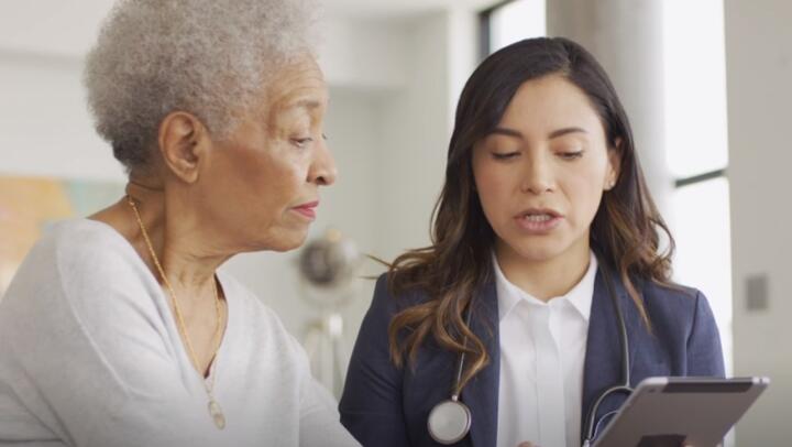
Your radiologist will look at your ultrasound images and provide your doctor with a diagnosis based on the ultrasound images.
How is a carotid ultrasound performed?
Your carotid ultrasound will be performed in a hospital or outpatient setting. The procedure takes less than an hour and generally includes these steps:
You will dress in a patient gown or wear your own loose-fitting, open-necked clothing. You will need to remove all jewelry around your neck area.
You will lie on your back on a procedure table. The radiologic technologist may move or tilt the table during your exam.
Your radiologic technologist will put ultrasound gel on your neck. The gel helps the ultrasound equipment make full contact with your skin by eliminating air. It also allows the equipment to slide easily across your skin without discomfort.
Your radiologic technologist will then place an ultrasound transducer on your skin. The transducer is a handheld wand that sends and receives the sound waves. The radiologic technologist presses it firmly onto your skin and moves it around to see the arteries and surrounding tissues. The transducer and sound waves are painless.
You may have to move or tilt your head or shoulders to get the best angle for the transducer.
Once the exam is complete, your team will wipe off the gel. The gel is water-based and washes away easily.
You may wait a short period of time while the radiologic technologist or radiologist verifies that the imaging is complete. You will probably go home right after the exam.
Will I feel pain?
Your comfort and relaxation is important to you and your care team. A carotid ultrasound is generally a painless imaging procedure. If you are already tender or sore in the neck, you may have some discomfort caused by mild pressure from the transducer. Take a few long, deep breaths to help yourself relax. Tell your technologist if any discomfort does not pass quickly.
How do I prepare for my carotid ultrasound?
There is no preparation needed for a carotid ultrasound.
Questions to ask your doctor
Preparing for a carotid ultrasound can be stressful. It is common for patients to forget some of their questions during a brief doctor’s office visit. You may also think of other questions after your appointment. Contact your doctor with concerns and questions before a carotid ultrasound and between appointments.
It is a good idea to bring a list of questions to your appointments. Common questions include:
Why do I need a carotid ultrasound? Are there any other options for diagnosing or screening my condition?
How long will the procedure take? When can I go home?
When and how will I receive the results of my test?
What other tests or treatments might I need?
When should I follow-up with you?
When and how should I contact you? Ask for numbers to call during and after regular hours.
What can I expect after my carotid ultrasound?
Knowing what to expect after a carotid ultrasound can help you get back to your everyday life as soon as possible.
How will I feel after the carotid ultrasound?
Standard diagnostic or screening ultrasound exams are painless, noninvasive imaging procedures. Call your doctor if you have any pain or discomfort after the ultrasound. You should be able to return to your normal activities immediately after the carotid ultrasound.
When can I go home?
Patients usually go home immediately after imaging is complete.
When should I call my doctor?
It is important to keep your follow-up appointments after a carotid ultrasound. Contact your doctor for questions and concerns between appointments.